
When Diagnostic Ultrasound for Foot Pain Helps
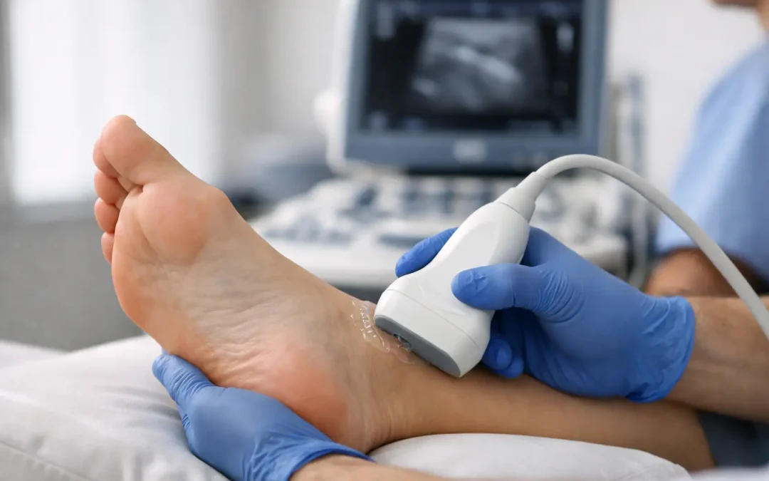
A sore heel that returns every morning, pain along the outside of the ankle on a run, or a forefoot that swells after walking can all have different causes. Diagnostic ultrasound for foot pain gives your podiatrist a live view of the soft tissues involved, helping to move beyond guesswork and towards a treatment plan that makes sense for you.
At Eclipse Foot Clinic, ultrasound is used as part of a wider specialist assessment. It is not simply about producing a scan image. The value comes from combining what the scan shows with your symptoms, medical history, footwear, activity levels, movement and hands-on examination.
What is diagnostic ultrasound for foot pain?
Diagnostic ultrasound uses high-frequency sound waves to create images of structures beneath the skin. In podiatry, it is particularly useful for assessing soft tissues, including tendons, ligaments, fascia, bursae, muscles and fluid-filled areas around joints.
A small handheld probe is moved over the area of concern with ultrasound gel applied to the skin. The images appear immediately on a screen, allowing your podiatrist to assess the painful structure while also asking you to move your foot, stand, or point to the exact area that hurts.
Unlike an X-ray, ultrasound does not use radiation. It is quick, comfortable and non-invasive. It can also be dynamic, meaning we can watch a tendon glide, assess a joint during movement, or compare the painful side with the other foot at the same appointment.
When an ultrasound scan may be useful
Foot pain is not always straightforward. A diagnosis based only on where pain is felt can be misleading, because several structures may sit close together. For example, heel pain may relate to the plantar fascia, a bursa, a nerve, a stress injury or a combination of factors affecting how you load the foot.
Ultrasound can be particularly helpful where pain is persistent, keeps returning, has not improved with sensible self-management, or is preventing you from exercising, working comfortably or wearing your usual footwear. It may also be recommended when swelling, a lump, thickening, clicking or a sudden change in symptoms needs closer assessment.
Common conditions ultrasound can help assess
Ultrasound is often used to investigate plantar heel pain, including suspected plantar fasciitis or plantar fascia thickening. It can identify changes within the fascia and help distinguish this from other soft-tissue causes of heel discomfort.
It is also valuable in tendon problems. This includes Achilles tendinopathy, peroneal tendon pain on the outer ankle, posterior tibial tendon problems on the inside of the ankle, and tendon irritation affecting the top or front of the foot. The scan may show tendon thickening, inflammation around the tendon sheath, small tears or changes in the tendon’s structure.
Other possible uses include assessing bursitis, joint swelling, synovitis, ganglion cysts, soft-tissue masses and some ligament injuries. For people with pain beneath the ball of the foot, ultrasound may assist with assessing the tissues around the metatarsal heads and identifying features that could be consistent with a neuroma or bursal irritation.
The scan is not used in isolation to label every finding as a problem. Tendon and fascia changes can occasionally be seen in people without pain, especially as we get older or remain very active. Your symptoms and clinical examination still matter.
What happens during your appointment?
Your appointment starts with a conversation about the problem: when it began, what brings it on, whether there was an injury, what footwear you use and how the pain affects your day. If you run, play sport or spend long periods on your feet at work, we will want to understand changes in training, surfaces, mileage and loading.
Your podiatrist will then examine the foot and lower limb. This may include checking range of movement, strength, joint mobility, swelling and tenderness, as well as watching how you stand and walk. Where appropriate, gait analysis can add useful information about the way forces travel through your feet and legs.
During the scan, the probe is placed over the relevant area. You may be asked to move your foot or ankle, contract a muscle, or stand briefly so we can assess the structure under load. Most scans take only a short time, and you can usually see the images as they are taken.
The real benefit is that findings can be discussed there and then in plain English. You should leave with a clearer understanding of what may be causing your symptoms, what is less likely, and which next steps are appropriate.
Ultrasound is part of the diagnosis, not the whole diagnosis
A scan is powerful, but it does have limits. Ultrasound is excellent for many soft tissues close to the skin, yet it does not show every problem equally well. Bones, deep joint structures and some stress injuries may require other investigations, such as an X-ray or MRI, depending on the clinical picture.
Equally, a normal-looking scan does not mean pain is not real. Nerve sensitivity, joint mechanics, referred pain and loading issues can all contribute to symptoms without producing one obvious image finding. This is why an experienced podiatric assessment remains essential.
If there is concern about a fracture, infection, significant circulation issue or another condition needing urgent medical input, you may be advised to seek further investigation or referred to the most appropriate service. Good diagnosis means recognising when ultrasound is the right tool and when it is not.
How scan findings can guide treatment
The purpose of imaging is to make treatment more precise. If a scan supports plantar fascia irritation, for instance, your plan may focus on managing pain, improving calf and foot function, adjusting activity, reviewing footwear and considering orthotic support where it is clinically appropriate.
For tendon problems, treatment commonly centres on carefully progressed strengthening, managing training load and addressing movement or footwear factors that may be keeping the tendon under strain. Some people benefit from shockwave therapy, while others need a period of relative rest, a change in footwear, an insole adjustment or a more structured rehabilitation programme.
Where inflammation is clearly identified and conservative care has not delivered enough improvement, injection therapy may be discussed. This is never a one-size-fits-all decision. The location of the problem, the tissue involved, your health history, goals and the potential risks all need proper consideration.
For active patients, the answer is rarely to stop everything indefinitely. It is usually about finding the right level of activity while the painful tissue settles and becomes more capable of handling load. For those whose pain affects everyday walking, the focus may be on restoring comfort, confidence and mobility as quickly and safely as possible.
When not to wait with foot pain
Book an assessment promptly if pain is worsening, you cannot comfortably bear weight, or there is marked swelling after an injury. Urgent medical advice is also needed for a hot, red or rapidly swelling foot, a wound that is not healing, new numbness, or a painful foot in someone with diabetes.
A sudden pop at the back of the ankle, followed by weakness or difficulty pushing off, needs urgent assessment as it may indicate a significant Achilles injury. These symptoms should not be managed by simply trying to push through.
A clearer route back to comfortable movement
Persistent foot pain can make every step feel uncertain, particularly when you have already tried rest, new trainers or stretches without a lasting result. Diagnostic ultrasound can provide useful answers, but more importantly, it can help shape a treatment plan around the structure involved and the life you want to get back to.
Whether your goal is completing a parkrun, walking the dog without heel pain, returning to work in comfortable shoes or simply getting through the day with less discomfort, a focused assessment is a positive place to start.